


Fetal Anatomy Scan also known as 20-week ultrasound or anomaly scan is a prenatal ultrasonography that is usually performed between 18 and 22 weeks of the pregnancy. It is a level 2 ultrasound that helps the doctors properly evaluate the health condition of the baby.
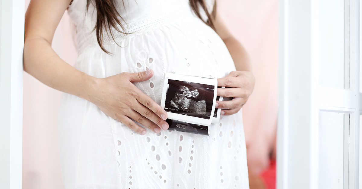
If you’re pregnant then you consult an experienced gynecologist in Siliguri so that he can guide you to do an anatomy scan. During this scan, you also witness your baby moving around your womb which is indeed a great experience. Additionally, this scan is also beneficial in detecting any fetal problems for early treatment.
Given below are the procedure and the purposes of the fetal anatomy scan.
Procedure
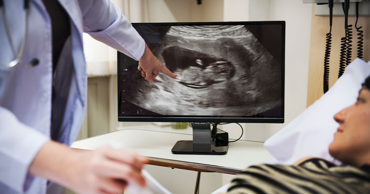
The doctor will direct you to lie down on an examination table and ultrasonic gel will be spread around your abdomen. This gel helps in making proper contact with the skin to pass the sound waves. The ultrasound technician will then move a handheld probe to various spots on the abdomen. This will help the technician in taking certain measurements of the fetus and by freezing the screen various images will also be taken for further analysis.
During this scan, the technician may draw some lines for measuring the fetus’s limbs and brain structure. You may also be directed to drink something or move a little to make the fetus move so that precise measurements can be taken. You can visit the best gynecologist hospital in Siliguri to get appropriate results from the scan.
Purposes
- Examines Face And Brain Defects
During the test, the technician will assess the shape of the back of the brain i.e., the cerebellum, and will also examine the spaces inside the brain. This helps in detecting the presence of any cysts in the choroid plexus. If there are any cysts in the brain areas then it may indicate chromosomal abnormalities. The defects in the face of the fetus such as cleft lip and palate can also be detected if the baby is in the right position.
- Check Maternal Conditions
The maternal conditions that are analyzed by the anatomy scan include checking the state of the cervix, uterus, and ovaries. It also assesses the position of the placenta to make sure that it is not in the cervix areas. If the placenta is covering the cervix then it may indicate placenta previa, which is lead to many dangers during the delivery. The blood flow in the umbilical cord and amniotic fluid amount are other significant measurements taken during the scan.
- Detects Congenital Defects In Heart And Spine
Any congenital heart defects can also be identified with the help of the scan which is one of the most common birth defects. The heart rate, positioning of the stomach and heart, and presence of four chambers and arteries are some of the factors that help to determine the proper functioning of the heart. In addition, the alignment of the vertebrae and skin covering the spine are analyzed to detect any defects in the spinal cord.
Undergoing this scan by taking the guidance of a qualified gynecologist in Siliguri is extremely essential. This is because apart from the above-mentioned reasons, this scan also helps in assessing the condition of the baby’s bladder, kidney, diaphragm, and abdominal wall. The issues of congenital anomalies can be hugely reduced with this painless scan method.

