


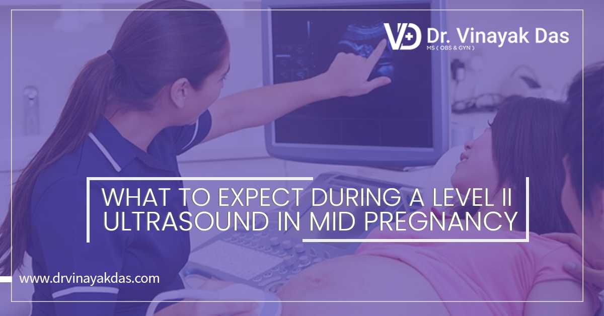
A level II ultrasound is a special test that pregnant women are asked to do that gives specific details about the growing baby. It is done for all moms-to-be to check how the baby is doing inside.
Today, it is very common for doctors to schedule a level 2 ultrasound, commonly called the 20-week anatomy scan. This is done because the test is a great way to see the development of the baby and offers reassurance that everything is going the right way for the baby and the mother.
Table of content:
1. When is a level 2 ultrasound done?
2. What to expect from a level 2 ultrasound
3. Risks
When is a level 2 ultrasound done?
Most anatomy tests are normally performed in the second trimester of pregnancy, which is at 20 weeks but it can also be done between 18 weeks and 22 weeks. If the pregnancy is a twin or multiple then more than one ultrasound is done.
What to expect from a level 2 ultrasound?
Between 18 weeks and 22 weeks, the baby is large enough for the medical personnel to see the baby's development which includes details of organs and limbs from head to toe in an anatomy scan.
If the woman is carrying multiples, each fetus will have its own scan. Follow-up ultrasounds are done to re-evaluate and keep a check on the growth and development of the baby.
Preventing any specific high-risk issues, the test examines the following regions to rule out any birth defects and other anomalies:
· Evaluation of the brain including ventricles, cerebellum, corpus callosum, and other key structures
· Neck including nuchal fold thickness
· Face structure including palate, eyes, nose, lips, and ears
· Detailed heart and lung anatomy
· Spine and ribs
· Abdominal organs like the stomach, intestines, spleen, liver, and the abdominal wall
· Limbs and digits
· If genitals are visible it determines the sex of the fetus
· Umbilical cord including vessels and insertion sites
· Placenta structure and location
· Cervical length
· Fetal position and movements
· Amniotic fluid
The baby will be measured from crown to rump, around the middle, and the head and the weight of the baby will be estimated. At level 2 checks are done for hard or soft markers, characteristics that may indicate an increased risk of a chromosomal abnormality.
Risks
There are no such risks known to be associated with ultrasound, however, unnecessary exposure to them is not advisable. This is why a handful number of ultrasounds within specific time frames are scheduled during pregnancy.
You can always to your medical personnel about what they are looking for and how the baby looks to them. Once they explain it, it will be helpful and knowledgeable for you.